Cellular and molecular biology

Presentation

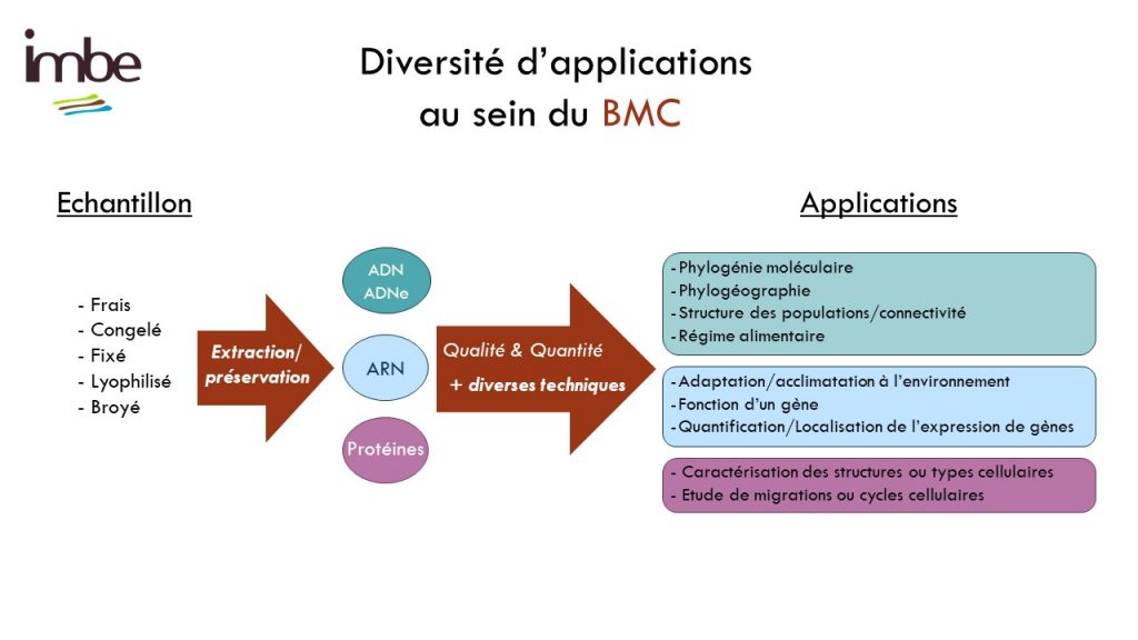
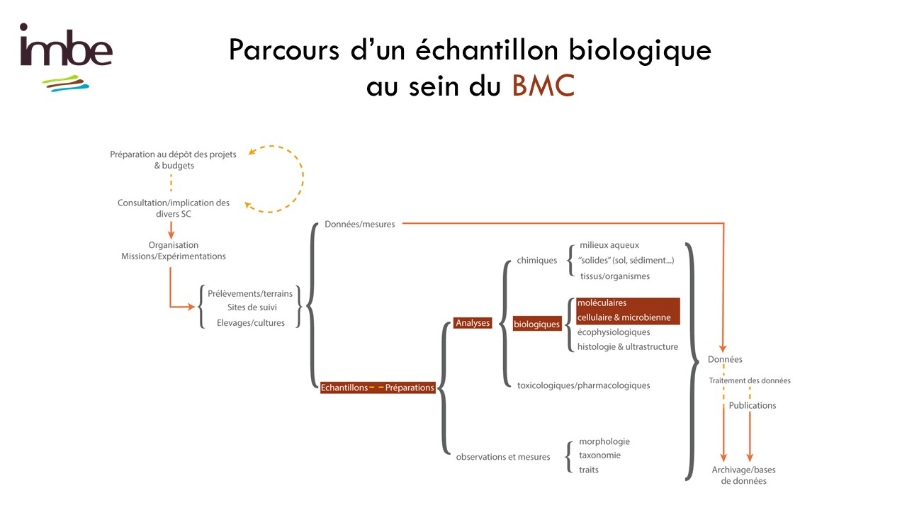
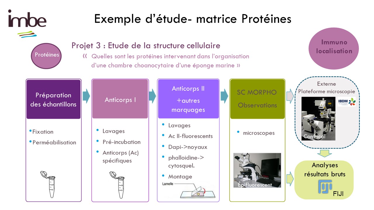
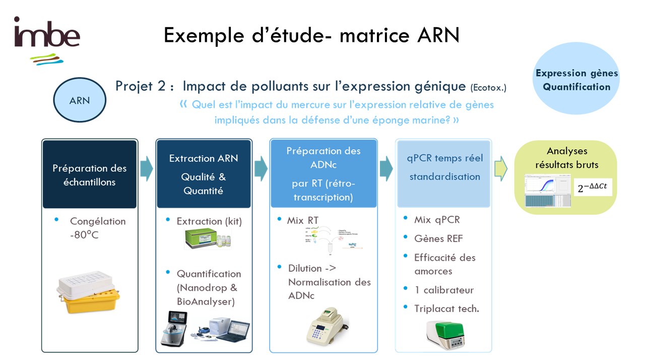
Le molecular and cellular biology department (BMC) is a technical platform located at the Endoume Marine Station, dedicated to the development of research projects focusing on the study of living macromolecules (DNA, RNA and proteins). It covers 250m2 of experimental laboratory space, with 15 shared rooms, each dedicated to a different type of experiment. In order to ensure a quality approach, a coherent 'one-way' circulation system (walking forward type) has also been put in place to limit the risks of sample contamination. This configuration is particularly advantageous for sensitive approaches such as metabarcoding.

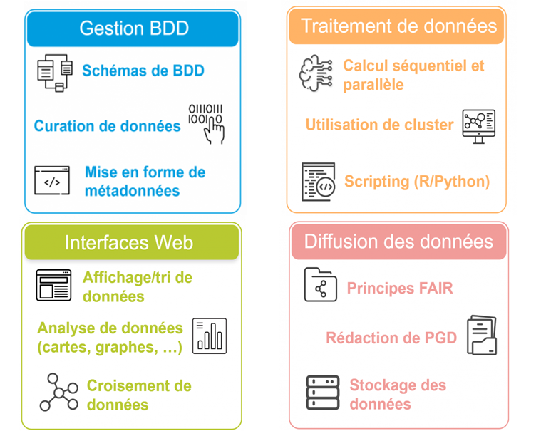
The BMC's technical platform offers technical support services, training and advice to researchers and their teams, thanks to the support of the service team. In addition, the variety of shared equipment and consumables available enables the BMC to provide scientific leverage to tackle complex technical challenges while meeting the needs of the unit's thematic diversity. This includes the study of numerous unconventional biological models, whether marine or terrestrial, animal or plant.

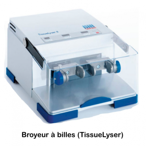
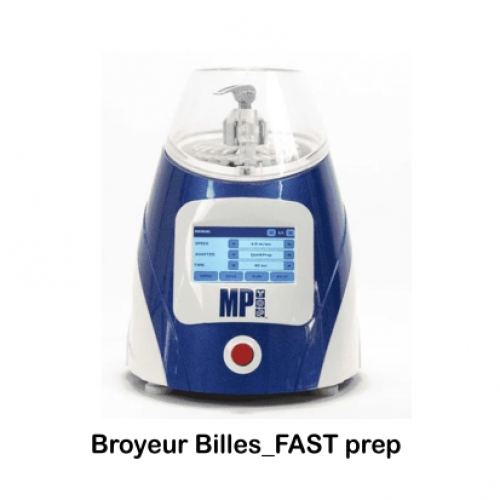
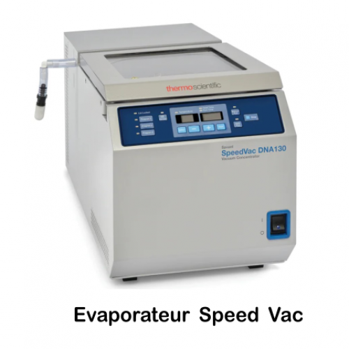
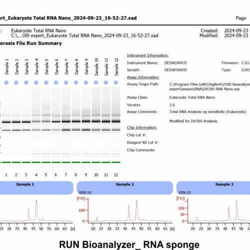
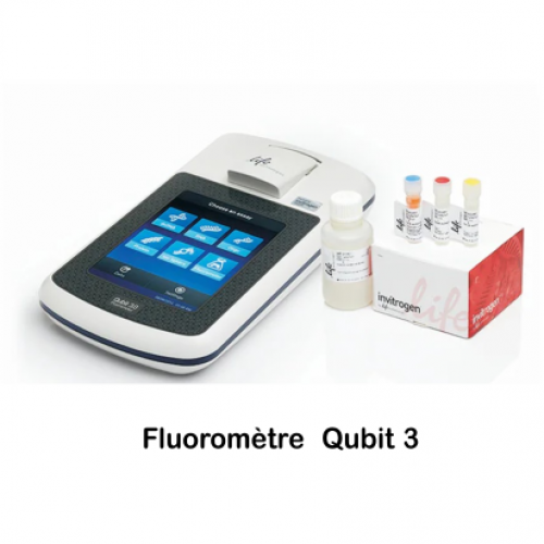
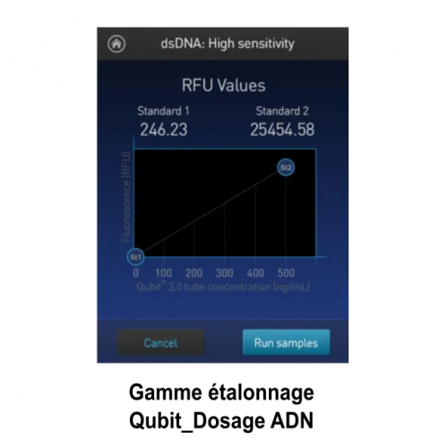
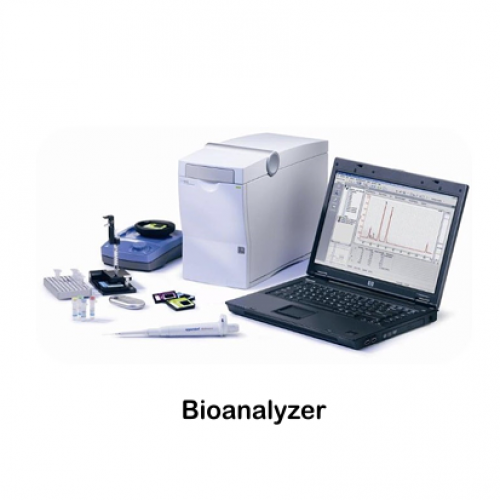
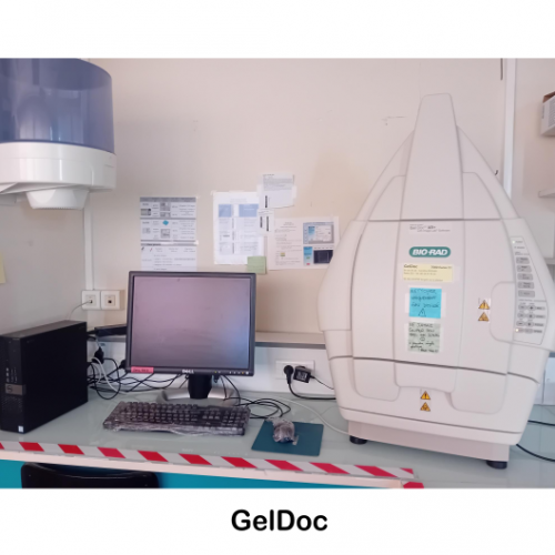
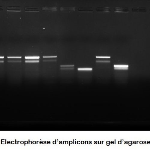
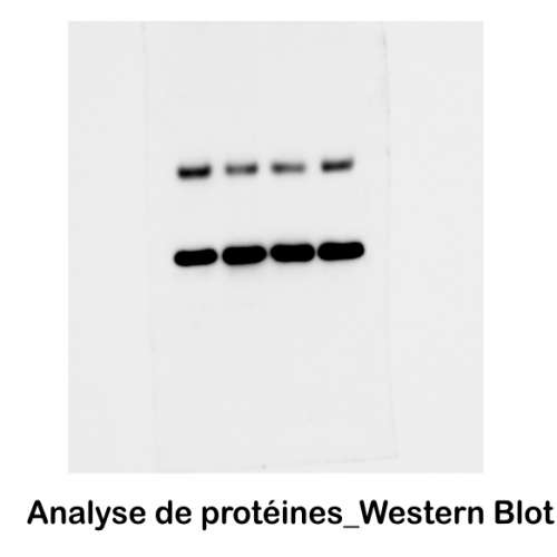
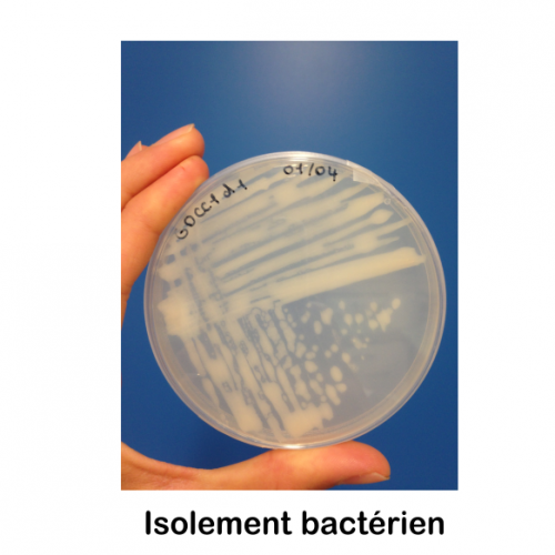
Instruments and techniques

The Team

Scientific manager of the BMC department
MCF-Maitre de Conferences
Technical manager of the BMC department
ITA-Engineer Administrative Technician
Search for a team member
Cécile Chemin
ITA-Engineer Administrative Technician
Caroline Rocher
ITA-Engineer Administrative Technician
Quentin Schenkelaars
MCF-Maitre de Conferences

Photo gallery

  • The equipment available enables different types of samples (soil, tissue, culture, etc.) to be prepared (weighed, dried, ground, dissociated, fixed).
  • Various types of macromolecules (gDNA, eDNA, RNA, Proteins) can be extracted from prepared samples.
  • Nanodrop and Qubit are used to quantify and assess the quality of extracted nucleic acids and proteins. The Nanodrop measures the concentration and purity of samples, while the Qubit offers a sensitive method for estimating the quantity of DNA, RNA or protein. Together, these tools guarantee accurate and reliable results, which are essential for subsequent analyses. This approach will help to optimise experimental protocols in molecular biology.
  • The Agilent 2100 Bioanalyzer system is a well-established automated electrophoresis solution for quality control of biomolecule samples. The system includes an instrument, data processing software, reagents and a dedicated microfluidic chip for DNA, RNA or protein analysis. It is suitable for next-generation sequencing (NGS), gene expression, biopharmaceutical and genome editing workflows, offering highly accurate analytical evaluation of various sample types.
  • PCR (polymerase chain reaction) is used to amplify specific DNA sequences using thermal cyclers, making them easier to study. After amplification, the products are analysed by gel electrophoresis, where the DNA fragments migrate according to their size. Finally, gel imaging (Gel Doc XR+) captures the image of the DNA bands, making it easier to interpret the results. PCR products can also be inserted into plasmids and then cloned into commercial bacteria. E.coli type I by transformation.
  • SDS-PAGE (polyacrylamide gel electrophoresis) is a technique for separating proteins based on their size, enabling their composition to be analysed. Western blotting is then used to transfer these proteins to a membrane and detect them specifically using antibodies. The proteins are visualised using a Chemidoc system, which uses chemiluminescence methods to capture images. These combined techniques enable the expression and modification of proteins in various biological samples to be studied.
  • Hybridization in situ (HIS) is a technique used in molecular biology to locate and visualise DNA or RNA sequences directly in cells or tissues of whole individuals, without removing them. This method makes it possible to observe gene expression in its spatial context, providing valuable information about development, differentiation and cellular interactions. By using labelled probes, it is possible to detect specific genes and study their localisation and regulation. This approach is particularly useful in fundamental biological studies. Manual HIS can be automated using the InsituPro VSI robot.


  • Immunohistochemistry (IHC) are techniques capable of detecting the presence of proteins in tissues using specific antibodies followed by secondary antibodies labelled with a fluorophore. They are essential for studying the localisation of proteins in fixed tissues. Manual IHC can be automated using the InsituPro VSI robot.


  • qPCR or quantitative PCR is a molecular biology technique used to amplify and quantify a specific DNA sequence or cDNA in real time. Unlike conventional PCR, it measures the quantity of DNA or cDNA in each cycle, thanks to the emission of a fluorescence that is proportional to the amplification. Its efficiency and precision make it an essential tool for comparing gene expression or the number of copies of a target sequence in samples.


  • Electroporation is a technique used to introduce molecules such as plasmids, RNAi and morpholinos into cells or living organisms. It involves applying an electric field to create temporary pores in the cell membrane, allowing these molecules to enter. For example, this method is essential for studying gene expression or protein function.

The BMC is sectorised to limit the risk of contamination reagents and samples with exogenous DNA and amplicons. The flow between sectors 1, 2 and 3 is monodirectional, or "forward".